Case Western Reserve University researchers and their collaborators in England have taken the first image of an open K+ channel — the beginning of a better understanding of how and why heart and nerve drugs work for some people and not for others.
The image and its analysis — featured in the July issue of the journal Structure — could some day help drug developers, contract research organizations and regulators develop safer, more effective drugs for cardiac and neurological diseases.
Mark Chance, director of the Case Western Reserve University School of Medicine’s Center for Proteomics and Bioinformatics, and Sayan Gupta, instructor at the center, worked with a research team at the University of Oxford to take the first high-resolution picture of the open state of a K+ channel, the university said in a release.

Using Informed Awareness to Transform Care Coordination and Improve the Clinical and Patient Experience
This eBook, in collaboration with Care Logistics, details how hospitals and health systems can facilitate more effective decision-making by operationalizing elevated awareness.
The picture shows the structure of an open channel — a pore-forming protein that allows potassium into a cell — which is enabling the researchers to analyze gating mechanisms that are important to heart function and nerve signaling.
And that is giving Case Western Reserve investigators a deeper understanding of G-protein coupled receptors (GPCR), which are involved in many diseases and are targets for more than half of today’s drugs.
“These receptors are key to the rhythm of your heart beating and your heart continuing to beat properly,” said Chance, who also is a professor of physiology and biophysics at the university and who has for years been developing biotechnology methods to study GPCRs.
Chance and the proteomics center have received a $1.1 million, four-year grant from the National Institute of Biomedical Imaging and Bioengineering to further study the receptors, in collaboration with Krzysztof Palczewski, chair of the university’s Pharmacology Department, and Gupta, who works at the Case Center for Synchrotron Biosciences at the Brookhaven National Laboratory in Upton, New York.
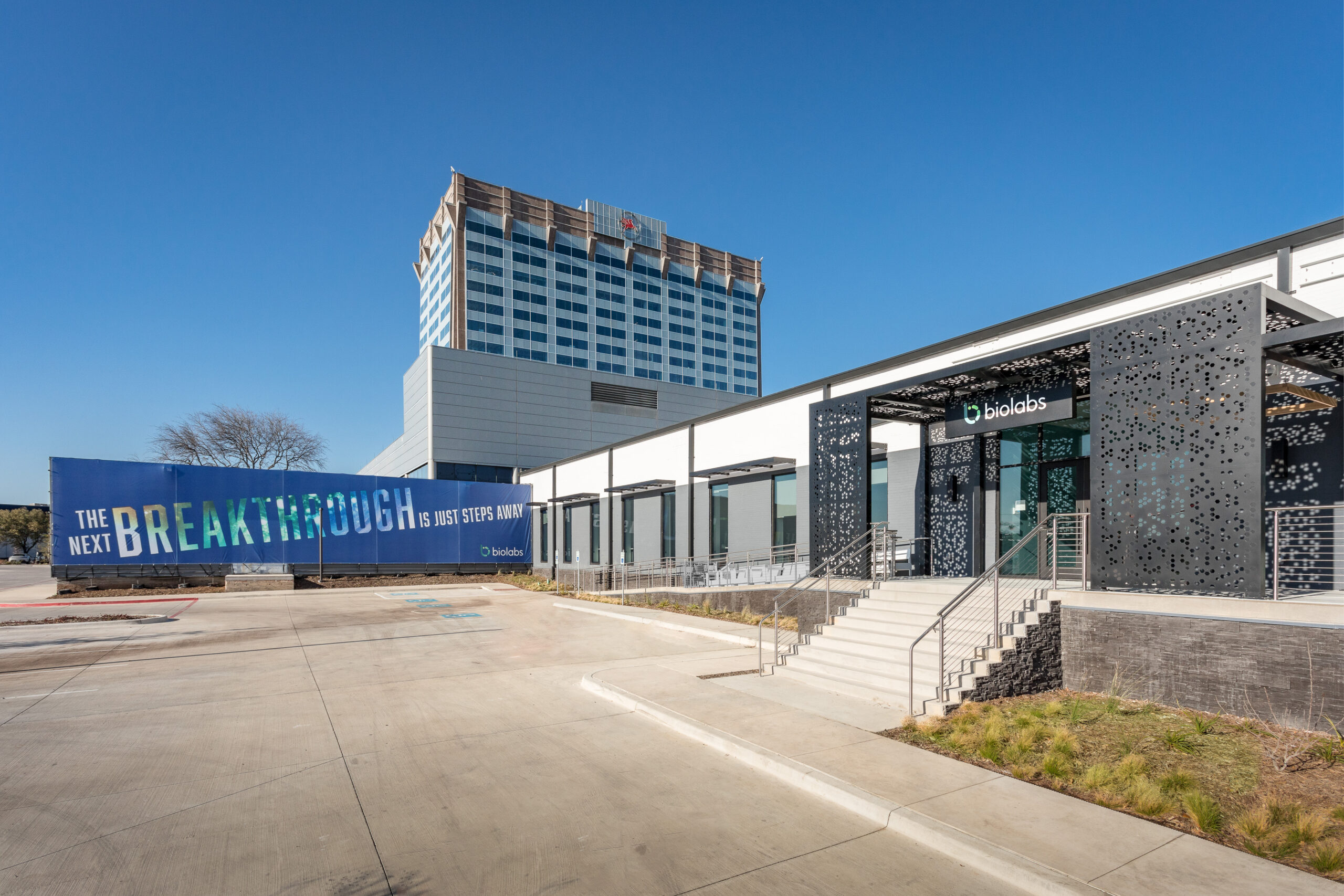
BioLabs Pegasus Park Cultivates Life Science Ecosystem
Gabby Everett, the site director for BioLabs Pegasus Park, offered a tour of the space and shared some examples of why early-stage life science companies should choose North Texas.
For their image, Chance and Gupta created a bacteria model of the K+ channel, which goes one way — in but not out. “Channels are very important to heart drugs that people take and are related to calcium blockers,” Chance said. “To understand how these drugs interact with the ion channels and regulate their flow of ions is a very important problem.”
To develop drugs, you have to know the structure of your target, said Dr. Dan Simon, chief of Cardiovascular Medicine at University Hospitals Case Medical Center and director of the Case Cardiovascular Center. “So the problem with the field up until now has been these ion channels have only had crystal structure in their closed form.”
Seeing the open form for the first time could teach scientists how drugs — like anti-arrhythmic drugs for the heart — work. “It paves the way to develop more rational drugs, and potentially, both more effective and safer drugs,” said Simon, who also is the Herman K. Hellerstein Professor of Cardiovascular Research at Case Western Reserve School of Medicine.