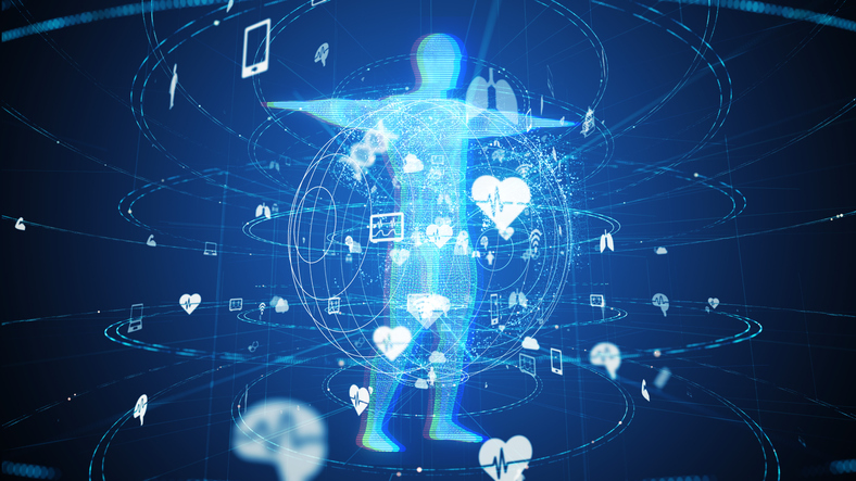
We can now grow complex human brain tissue in a test tube.
Scientists in Vienna, Austria, used a method that allows pluripotent stem cells to develop into mini-brains (i.e., cerebral organoids) that even have several distinct brain regions, according to an article in this issue of Nature.
While this year has seen 3-D printed miniature human livers and even regenerated human heart tissue that beats on its own, this milestone at the Institute of Molecular Biotechnology of the Austrian Academy of Sciences is certainly a contender for most impressive yet. And, of course, it has big implications for pharma.
With the Rise of AI, What IP Disputes in Healthcare Are Likely to Emerge?
Munck Wilson Mandala Partner Greg Howison shared his perspective on some of the legal ramifications around AI, IP, connected devices and the data they generate, in response to emailed questions.
“In addition to the potential for new insights into the development of human brain disorders, mini-brains will also be of great interest to the pharmaceutical and chemical industry,” Dr. Madeline A. Lancaster, research team member and first author of the publication, said in a release. “They allow for the testing of therapies against brain defects and other neuronal disorders. Furthermore, they will enable the analysis of the effects that specific chemicals have on brain development.”
To create the tissue, they started with human embryonic stem cell lines and induced pluripotent stem cells. “We modified an established approach to generate so-called neuroectoderm, a cell layer from which the nervous system derives. Fragments of this tissue were then maintained in a 3-D culture and embedded in droplets of a specific gel that provided a scaffold for complex tissue growth. In order to enhance nutrient absorption, we later transferred the gel droplets to a spinning bioreactor. Within three to four weeks, defined brain regions were formed,” Dr. Jurgen Knoblich, who led the research team at the Institute of Molecular Biotechnology, said in a release (emphasis added).
According to the release (emphasis added):
Already after 15 – 20 days, so-called “cerebral organoids” formed [that] consisted of continuous tissue (neuroepithelia) surrounding a fluid-filled cavity that was reminiscent of a cerebral ventricle. After 20 – 30 days, defined brain regions, including a cerebral cortex, retina, meninges as well as choroid plexus, developed. After two months, the mini-brains reached a maximum size, but they could survive indefinitely (currently up to 10 months) in the spinning bioreactor. Further growth, however, was not achieved, most likely due to the lack of a circulation system and hence a lack of nutrients and oxygen at the core of the mini-brains.
A Personalized Approach to Medication Nonadherence
At the Abarca Forward conference earlier this year, George Van Antwerp, managing director at Deloitte, discussed how social determinants of health and a personalized member experience can improve medication adherence and health outcomes.
Using these mini-brains, the scientists were able to model a neuronal development disorder in which brain size is significantly reduced — microcephaly — and note that a change in the direction the stem cells divide might cause the disorder.
The brains could, of course, be used to learn more about other disorders. But where else could this lead? Testing implantables on mini-brains instead of animals first? Faster and more accurate results in trials?
Leave your thoughts about what this could mean for pharma, the medical device industry or medicine in the comments below.